|
ULTRASOUND OF
PLACENTAL INFARCTION |
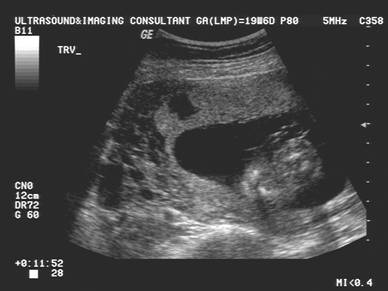
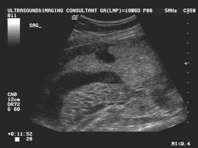
- Infarct is not sonographically visible if the villous structures remain preserved without increased cellularity.
- Increased cellularity results in hyperechoic areas (1).
- Fibrin deposition results in hypoechoic areas (1).
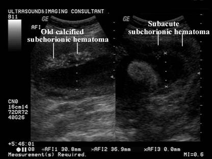
- Old infarcts are cystic or hypoechoic and is indistinguishable from decidual cysts or intervillous thrombus.
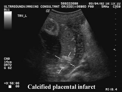
- Calcification (very rare) (2).
|
|
|
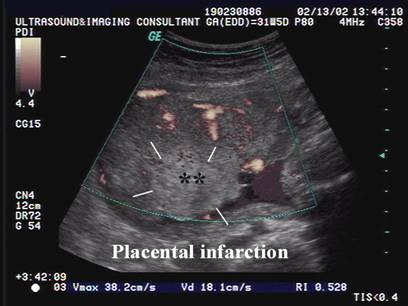
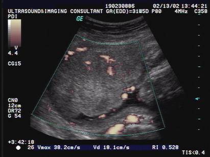
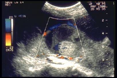
- Color doppler may detect areas of placental ischemia or infarction by the relative absence of blood flow in the affected area (3).
|
|
|
|
|
|
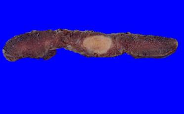
- A rare type of infarction involves massive deposition of fibrin in the basal plate of the placenta causing engulfment of the adjacent villi that may become avascular and atrophic. In the largest study (4), the hypoechoic areas that corresponded to the infarction were identified in four placentas. Twelve of thirteen cases showed fetal growth restriction while eight fetuses were stillborn.
|
|
|
|
|
|
REFERENCES |
- Sherer DM, Allen TA, Metlay LA et.al. Linear calcification in a placental infarct causing complete distal sonographic shadowing. J Clin Ultrasound 1994;22:212-213.
- Bude RO, van de Ven CJ, Bowerman RA et.al. Evaluation of normal placental vasculature with with power doppler ultrasound. Radiology 1994;193:231.